Arthroscopic chondroplasty
Arthroscopic chondroplasty is a surgical procedure used to clean and smooth damaged cartilage in the knee. This minimally invasive procedure uses a small camera, known as an arthroscope, to look inside and small instruments to repair the knee. By using a small camera and instruments, the incisions can be small, resulting in a faster and less painful recovery.
Anatomy
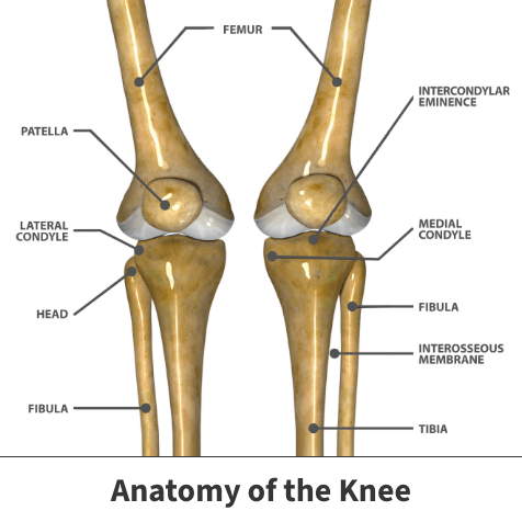
The knee is a hinge joint, meaning it can only bend in one direction. The knee is where the tibia, the femur and the patella meet. Like most joints in the body, the knee has a dense, fibrous, connective tissue, known as articular cartilage, that seals the joint space between the femur and tibia. This cartilage prevents the bones from rubbing against each other. It also acts as a shock absorber and allows for smooth and stable movement. The knee has three major compartments:
- Medial compartment (the inside part of the knee)
- Lateral compartment (the outside part)
- Patellofemoral compartment (the front of the knee between the kneecap and thighbone)
About
Arthroscopic chondroplasty is a procedure used to repair small areas of damaged cartilage in the knee. The goal of surgery is to lessen friction in the joint, allowing the knee to move freely and without pain. Arthroscopy is minimally invasive surgical procedure. Conventional surgical procedures typically involve larger incisions, but arthroscopic procedures use smaller incisions. The incision size is not as big because your surgeon uses an arthroscope (small camera) to see what is happening during the surgery.
Symptoms
Arthroscopic chondroplasty treats damaged articular cartilage in the knee so that new healthy tissue can grow and allow the knee to move smoothly again. The procedure treats damage caused by trauma to the knee or degenerative conditions such as arthritis.
The procedure treats a variety of symptoms, including:
- Knee joint pain
- Stability issues
- Popping, locking, or “giving” of the knee
Treatment
First, the knee is positioned for the procedure, and the skin is cleaned and sterilized. Next, two to five small incisions are created around the front of the knee. An arthroscopic camera is inserted through one of the incisions. The other incisions are used as access points for other arthroscopic tools.
Once everything is ready to go, fluid is pumped in to expand the joint, which gives the surgeon a clear view, as well as room to work. The surgeon inserts small surgical tools to carefully remove damaged cartilage and any loose tissue.
After the knee repair, excess fluid is drained, the instruments are removed, and the incisions are closed. As the knee heals, new “scar tissue” cartilage grows over the bare spot to replace the missing cartilage.
Recovery
Recovery is fast, with relief felt almost immediately. There is little risk, scarring, or pain involved with this procedure.
Videos
Related specialties
- ACL Injuries
- Articular Cartilage Restoration
- Deep Thigh Bruising
- Fractures of the Tibial Spine
- Iliotibial Band Syndrome
- Lateral Collateral Ligament (LCL) Injuries
- MACI
- Medial Collateral Ligament Injuries
- Meniscus Tears
- Muscle Spasms
- Muscle Strains of the Calf
- Partial Knee Replacement
- Patellar Fracture
- Quadriceps Tendon Tear
- Runner's Knee
- Senior Strong
- Shin Splints
- Total Knee Replacement Surgery