Hip dysplasia
Dislocated hip
A hip dislocation is when the thighbone bone gets dislodged from the hip socket either forward or backward. This injury is caused by something with a high impact, like a fall or a car accident. The injury itself is extremely painful and is fixed by manually putting the thighbone back into place. Recovery takes a few months but generally has excellent results.
Anatomy

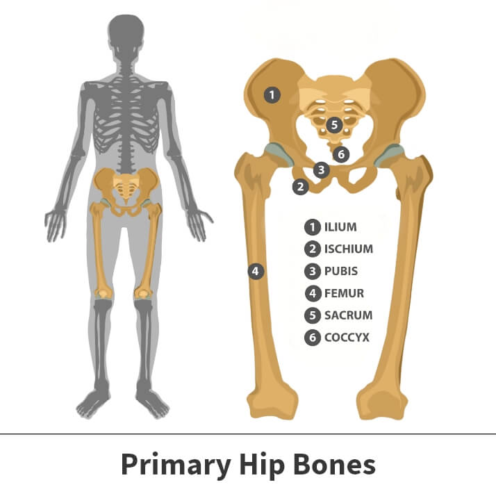
The hip is a ball and socket joint. The socket is formed by the large pelvis bone (acetabulum) and the ball is formed by the upper end of the thighbone (femoral head). A slippery tissue known as the articular cartilage covers the surface of the joint, creating a smooth surface that allows for the bones to glide easily across each other.
The large pelvis bone is ringed by strong fibrocartilage, known as the labrum, which forms a lining around the socket.
Additionally, the whole joint is surrounded by bands of tissue (ligaments) that form a capsule that hold the joint together. The undersurface of the capsule is lined by a thin membrane (synovium) which produces synovial fluid that lubricates the hip joint.
About
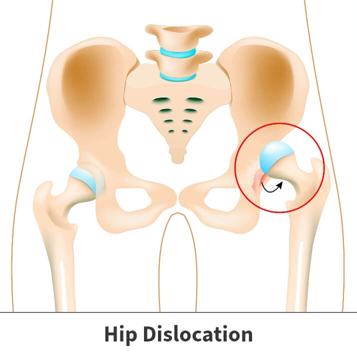
When the upper end of the thighbone is pushed out of the hip socket, it is known as a hip dislocation. The general cause of hip dislocation is impact injuries, such as car accidents and falls from significant heights.
There are two different types of hip dislocations – posterior dislocation and anterior dislocation.
Posterior dislocation – is the more common of the two types of hip dislocations and happens when the thighbone is pushed backward out of the socket. This type of hip dislocation will leave the lower leg in a fixed position with the foot and knee rotated inward toward the middle of the body.
Anterior dislocation – is less common and happens when the thighbone slips forward out of its socket, resulting in the hip being slightly bent and the leg being rotated out and away from the middle of the body.
Additionally, when the hip is dislocated, the ligaments, labrum, muscles, and other soft tissues holding the joint together are often damaged. The nerves around the hip may also be injured.

Symptoms
Hip dislocations are very painful and will result in you being unable to move your leg. If the injury results in nerve damage, you may not have any feeling in the foot or ankle area.
Diagnosis
Your Florida Orthopaedic Institute physician will take a look at your symptoms and general health. Hip dislocations can in most cases be diagnosed just by looking at the position of the leg. Since hip dislocations often occur along with additional injuries, your physician will perform a thorough evaluation to determine the full extent of your injury.
Your doctor may order imaging tests, such as X-rays, to show the exact position of the dislocated bones, as well as any additional fractures in the hip or thighbone.
Treatment
If there are no other injuries present, then your physician will give you an anesthetic or a sedative and manually readjust the bones back into their correct positions. This procedure is known as a reduction.
In rare cases, torn soft tissues or small bony fragments block the bone from going back into the socket. When this happens, surgery will be needed to remove the loose tissues and correctly position the bones.
Videos
Related specialties
- Anterior Hip Replacement
- Avascular Necrosis (Osteonecrosis)
- Groin Strains & Pulls
- Hamstring Injuries
- Hip Arthroplasty
- Hip Arthroscopy
- Hip Flexor Strains
- Hip Fractures
- Hip Hemiarthroplasty
- Hip Impingement Labral Tears
- Hip Muscle Strains
- Hip Pointers & Trochanteric Bursitis
- Iliopsoas Tenotomy
- Labral Tears of the Hip (Acetabular Labrum Tears)
- Osteoarthritis of the Hip
- Osteoporosis
- Pelvic Ring Fractures
- Piriformis Syndrome
- Sports Hernias (Athletic Pubalgia)
- Thigh Fractures
- Thigh Muscle Strains
