Diagnostic imaging
State-of-the-art musculoskeletal scans from orthopedic specialists
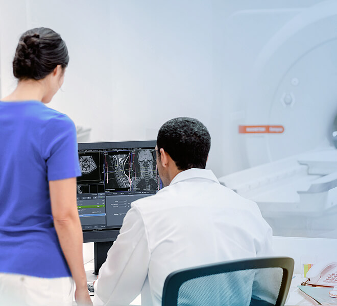
At Florida Orthopaedic Institute, you can have your diagnostic testing performed in the same facility as you receive your orthopedic care. Our highly skilled healthcare professionals use the most advanced technology to meet your medical needs.
Conditions that require diagnostic imaging
If our physicians suspect that you have one of the following injuries or conditions, they will ask you to schedule imaging so they can give you a proper diagnosis and treatment plan:
- Abnormalities in the Body
- Arthritis
- Back & Spine Pain
- Cysts
- Injuries or Abnormalities of the Joints
- Musculoskeletal Problems
- Painful or Injured Joints
- Slipped Discs
- Soft Tissue & Bone Pathology
- Spinal Conditions & Injuries
- Sports Injuries
- Tumors
- Tumors or Inflammation of the Spine
- Vertebrae of Intervertebral Discs of the Spine
Orthopedic diagnostic imaging
Our nationally renowned team of orthopedic specialists uses leading technology to meet your medical needs, including the following diagnostic imaging services:
A CT scan (sometimes called a “CAT scan”) is a simple, non-invasive procedure that enables medical professionals to quickly and safely obtain sophisticated and accurate diagnostic information on your brain, chest, abdomen, pelvis, spine and extremities. A CT scan can be up to 100 times more sensitive than conventional X-rays, allowing radiologists to distinguish subtle differences in soft tissues that may not be detected with regular X-rays.
An electromyogram (EMG) measures the electrical activity of muscles at rest and during contraction. Nerve conduction studies (NCS) measure how well and how fast the nerves can send electrical signals. If you have leg pain or numbness, these tests can help find out how much your nerves are being affected. They check how well your spinal cord, nerve roots, and nerves and muscles that control your legs are working. EMGs are done to find diseases that damage muscle tissue, nerves, or the junctions between nerve and muscle. Florida Orthopaedic Institute is one of the few facilities in the country accredited by the American Association of Neuromuscular & Electrodiagnostic Medicine (AANEM). The AANEM is the only EMG accrediting association in the country and recognizes facilities for achieving and maintaining the highest level of quality, performance, and integrity based on professional standards, and is structured to assess, evaluate, and improve patient care. It is the recognized standard for EDX medicine.
Fluoroscopy is a study of moving body structures – similar to an X-ray “movie.” A continuous X-ray beam is passed through the body part being examined. The beam is transmitted to a TV-like monitor so that the body part and its motion can be seen in detail. As an imaging tool, fluoroscopy enables physicians to look at many body systems, including the skeletal, digestive, urinary, respiratory and reproductive systems. Fluoroscopy also allows physicians to check anatomical structures in perfect alignment.
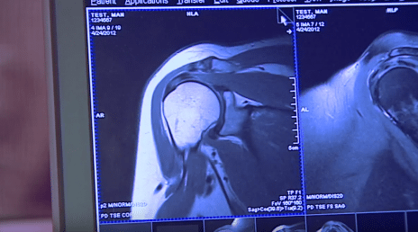
An MRI is an imaging test that allows physicians to visualize internal structures of the body for disease or abnormal conditions. MRI does not use radiation for imaging, like an X-ray or computerized tomography (CT) scan. Instead, MRI examinations use specialized equipment with a powerful, constant magnetic field, rapidly changing local magnetic fields, radiofrequency energy and dedicated equipment, including a powerful computer to create clear pictures of internal body structures. MRI examinations are performed in a special room that houses the MRI scanner. Patients lie down on a comfortably padded table for approximately 30 minutes to complete their scans. Once the entire MRI examination is complete, a report is generated for your physician to review to help determine treatment options.
For some MRI studies, a contrast agent is injected directly into the joint to better evaluate joint & soft tissue structures. This is often done first utilizing the guidance of the CT scanner and then completed in the MRI scanner. Arthrograms can be used for multiple joints including the shoulder, elbow, wrist, hip, knee & ankle. They are used to diagnose conditions that affect soft tissue in many areas of the body; such as, damaged ligaments, cartilage / joint injuries, rotator cuff tears, and disorders of the biceps tendon.
X-rays are one of the most common and widely available diagnostic imaging techniques and one of the oldest forms of medical imaging. An X-ray is actually an electromagnetic beam that is aimed through your body to a piece of film behind it. The resulting image allows your doctor to help narrow the causes of an injury or illness and ensure that the diagnosis is accurate.
“My first visit to Florida Orthopaedic Institute with Dr. Nydick was outstanding. Staff were unbelievable. Dr. Nydick took more X-rays. I would put the word ‘caring’ into all that I’ve experienced at the Florida Orthopaedic Institute, from the beginning to the end. If I wanted a copy of the X-ray, it was given to me. They 100% followed up on everything.” Vicki Kasper | Distal Radius Wrist Fracture